As a nurse working with patients, you’ll be responsible for assessing patients. When you start to work on specialty nursing units, you have to customize your assessment routine specifically for that patient population. Let’s discuss some important things to know for a cardiac assessment.

Table of Contents
3 Common Cardiac Issues
Before we get to tips about the cardiac assessment, you need to learn the three different issues that can happen with a person’s heart. If you understand these three things, it will make educating the patient easier and help you with your assessments.
The three types cardiac issues that normally arise typically involve:
- Electrical Abnormalities – the electrical system that tells the heart to beat.
- Structural Heart Problems – issues with the valves, septum, or large blood vessels carrying blood to and from the heart
- Coronary Artery Issues – blockages or spasms in vessels that supply the heart with blood (coronary artery blockages), which will compromise the heart’s ability to beat effectively
Your patient may have an issue with one or more of these, and often they overlap. However, it is very helpful to help predict complications and inform your patient education to get these separated out in your mind.
How To Do A Cardiac Assessment
Let’s look at how to do a thorough cardiac assessment. These are the exact steps I would take as a cardiac nurse after I get my report.
Cardiac Monitor
Before I head into my patient’s room, I look at what their heart is doing on the cardiac monitor. I compare that to the information I received from the off-going nurse and what’s documented in the chart to ensure it’s the same.
Vitals, Labs, and Medications
I look for anything that might impact their vitals signs. Some cardiac patients – especially ones that just had procedures will usually have blood pressure or heart rate parameters within which they are expected to fall. If their heart rate or blood pressure falls or jumps outside of the parameters, the physicians may have “as-needed” or PRN medications you can use. If not, you may need to notify them. I look for the trend of their vitals over the last shift or two – not just the most recent vitals. If any vitals were out of range, I look in the chart to see if any medications were given.
I also look for any cardiac-related medications I’ll have to give within the next hour or so, and what their normally scheduled ones are.
I also look for the potassium levels from the labs. Cardiac physicians always want to know what the potassium level is, and want it well within the 3.0-5.5 range. I also tend to look at whatever recent labs were drawn and take note of any abnormalities.
Observations
- At rest, do they seem short of breath? How hard are they breathing?
- What does their skin look like? Is it consistent with their ethnicity? Do they seem pale, ashen, blue, etc? Is it dry? Are they sweating?
- When you walk into the room, do they notice you? Can they turn their head, acknowledge you, and converse with you?
- Do they independently use and move all 4 extremities with ease, or is there a deficit?
- If you’re in an ICU and they’re on a monitor and asleep, note what their vitals are BEFORE you wake them, and again during your assessment to see if there is a large difference.
Head to Toe Cardiac-Focused Assessment
I first ask basic orientation questions: “Please tell me your name, where you are, what year it is, and why you’re here with me.”
Wish Your 12-Hour Shift Had A Map?👇
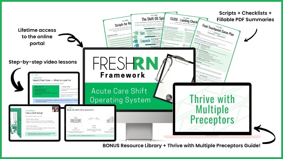
This course helps you turn a messy 12-hour acute care shift into a clear, structured rhythm. You’ll learn how to organize your day after report, prioritize when everything feels urgent, recover when something throws you off track, and give a stronger end-of-shift handoff. No fluff, no vague advice. Just a repeatable system you can use shift after shift. Designed specifically for med-surg, stepdown, and ICU nurses who are tired of feeling behind. No fluff. Just practical training you can use on your very next shift.
See What’s Inside Shift OS →
While they’re talking, I observe their face for equal movement and notice how they speak (if it’s clear, garbled, and so forth.) I also pay attention to how hard they’re working to breathe while speaking and if they can manage their secretions (are they choking on their spit?).
I have them smile to check facial symmetry. I have them look at me, hold their head steady and cover one eye, and then ask them to look directly at my nose. Then I hold up fingers in all four quadrants, one at a time, and see if they can tell me the correct number. Then, I do the other eye. This assesses visual quadrants and if they have a field cut.
Then, I see if they can follow my fingers with just their eyes. I hold their head steady by gently placing my index finger on their chin, then use my other hand to make a big X and see if they can follow it fully and equally with both eyes. I’m watching for nystagmus, and this exercise assesses their extraocular movements.
I’ll grab a penlight and shine a light in each eye (make sure you warn them first). I am looking at the pupil’s shape, size, and responsiveness to light.
Then I grab my stethoscope to listen to their lungs, heart, and bowels. I personally find it flows better to listen to posterior lung sounds (on their back) first. I will ask them to lean forward and listen to 8 spots on the back. (This is a great example of where, scroll down to BACK.) While listening, I’m visualizing their back. If they are bed bound, you can roll them to the side, but may need help. This would also be a great time to assess the coccyx of any bed bound patient if you do need to get them on their side to listen!
I then gently set them back on the bed and listen to lung sounds on the front. If they’re on an oxygen device, I’m observing what kind it is and noting if it’s hooked up correctly and working appropriately.
Then, I tell them to breathe normally so I can hear their heart.
Here is where I listen to their heart:
- The Aortic Valve – located at the second intercostal space right sternal border.
- The Pulmonary valve – located at the second intercostal space left sternal border.
- Erb’s Point – located at the third intercostal space left sternal border.
- The Tricuspid Valve – located at the fourth intercostal space at the left sternal border.
- The Mitral Valve – located at the fifth intercostal space midclavicular line
I listen for that good ol’ lub-dub, as well as for any odd sounds like a pericardial friction rub, murmurs, or additional heart sounds.
Finally, I move down to the abdomen and listen to each quadrant of their intestines to note the bowel sounds. Then, I casually flip my stethoscope back onto my neck and feel like such a badass every time I do. After, I gently palpate the abdomen to see if their is any distention or discomfort.
Then, I palpate radial pulses. After, I put my fingers in their hands and ask them to gently squeeze, then push and pull. At this time, I’m noting any skin issues and looking for any edema on the extremities. I’m also looking around at any IV insertion sites and the status of their dressings, as well as any devices that are inserted (chest tubes, arterial lines, central lines, dialysis catheters, feeding tubes, tracheostomies,
Finally, let’s work down to the feet. I feel pedal pulses (dorsalis pedis and posterior tib). If I can’t palpate them, I definitely get a portable doppler to do so. This is really important in cardiac patients! There is often edema in the feet, so I check that as well.
I ask about sensation and if they have any numbness or tingling anywhere. Many cardiac patients will say “Well, I always have that!” In response, I typically ask, “Is the numbness and tingling you’re feeling now different than your normal?”
For a motor assessment, I’ll then ask them to pick one leg up and hold it up for a few seconds, then the other one. I cap it off by having them push down on my hands (“Like you’re stepping on the gas pedal!”) and pull back up against my hands to check for dorsiflexion and extension strength and if it’s equal.
Final Thoughts About Cardiac Assessment
Whenever you perform a cardiac assessment, make sure you’re considering what you’re physically observing in context with their cardiac history and diagnostics. While it takes time to get comfortable assessing cardiac patients, you will be a pro at it in no time!
More Resources for Cardiac Assessment:
- 5-Lead ECG Interpretation, Electrocardiogram Tips for Nurses
- What is Telemetry and Telemetry Lead Placement?
- Nursing Management of Atrial Fibrillation
- Cardiac Assessment Checklist – free PDF download
Tired of Googling rhythms and hoping no one notices? 👀

Cardiac nursing can feel like you're drowning in alarms, rhythms, and unfamiliar meds before you even get report—and you're expected to keep up. But here's the truth: you don’t need to know everything, you just need to know what matters most.
Cardiac Confidence was created by experienced nurses to help you cut through the noise. We cover what actually shows up in real life—like post-cath care, recognizing unstable rhythms, caring for chest tubes, and knowing when to escalate (and a lot more).
No fluff, no advanced cardiac stuff you don’t need yet. Just focused, foundational info that makes a big difference.
Click for Instant Access


0 Comments