
Caring for patients on telemetry starts with understanding how the monitor works and placing the electrodes correctly. Telemetry monitors your patient’s heart rate and rhythm, giving you information to use alongside your bedside assessment.
This guide covers telemetry lead placement for a common five-electrode setup, skin preparation for a clear tracing, and the checks nurses should make when starting monitoring. We’ll also explain which patients may need telemetry and how it fits into everyday nursing care.
A quick note: This guide is for nursing education and does not replace clinical judgment, hands-on training, or patient-specific orders. Always follow your facility’s policies and your equipment manufacturer’s instructions. Read our full disclaimer.
What Is Telemetry?
In the hospital, cardiac telemetry continuously monitors a patient’s heart rate and rhythm. Electrodes (small adhesive patches on the skin) pick up the heart’s electrical signals. Wires connect the electrodes to a monitor or portable transmitter, allowing the care team to view the rhythm at the bedside or a central monitoring station.
Telemetry helps the team recognize heart rhythm changes, but it doesn’t tell you everything about how your patient is doing. You still need to assess their symptoms, vital signs, and overall condition. It also doesn’t replace a diagnostic 12-lead ECG when one is needed.
Telemetry Lead Placement
A common telemetry placement setup uses five electrodes: four on the torso and one at a selected chest position. Although we often call this “5-lead telemetry,” electrodes are the adhesive patches on the skin; leads are views of the heart’s electrical activity.
Where Do the Five Electrodes Go?
The diagram and table below show one common configuration of telemetry lead placement using the V1 chest position. Right and left refer to the patient’s right and left.
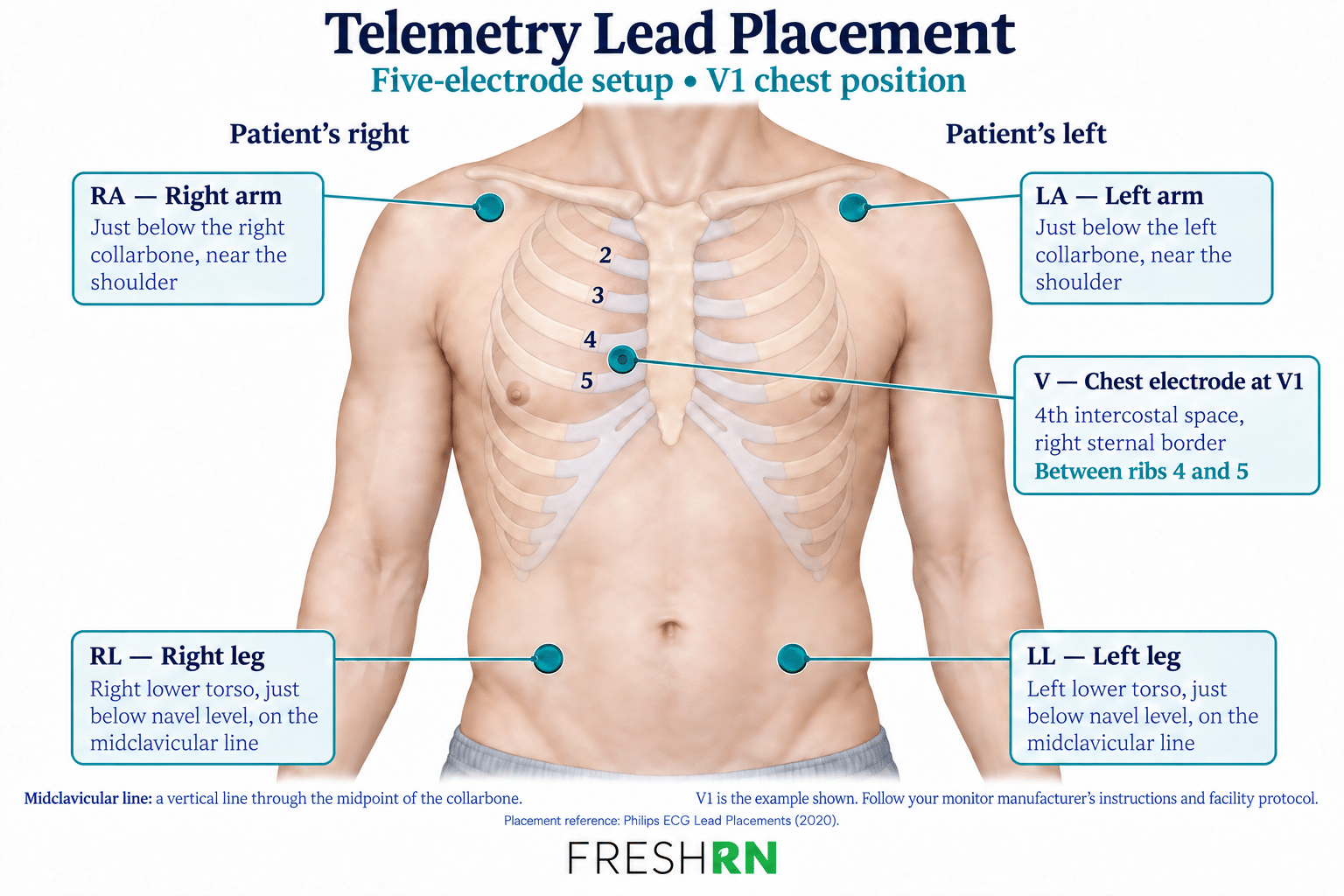
| Electrode label | Placement |
|---|---|
| RA — right arm | Just below the right collarbone, near the shoulder |
| LA — left arm | Just below the left collarbone, near the shoulder |
| RL — right leg | Right lower torso, just below the belly button’s level, along a vertical line through the midpoint of the collarbone |
| LL — left leg | Left lower torso, just below the belly button’s level, along a vertical line through the midpoint of the collarbone |
| V — chest electrode, positioned at V1 | Fourth intercostal space—the space between the fourth and fifth ribs—at the right edge of the sternum |
Follow your monitor manufacturer’s placement instructions and your facility’s protocol for telemetry lead placement. The chest electrode may use another designated position depending on the monitoring goal; V1 is the example shown here.
Verify the Order, Patient, and Telemetry Device
Before starting telemetry, confirm the monitoring order or applicable protocol, including the reason for monitoring. In an emergency, follow your facility’s emergency response protocol without delaying care to obtain a routine order.
Make sure the rhythm on the screen belongs to the patient you’re caring for:
- Use two patient identifiers, such as full name and date of birth or medical record number, according to facility policy. A room number is not a patient identifier.
- Match the telemetry device number to the patient’s monitoring record.
- Confirm the patient’s current unit and room with the monitoring team so they know where to direct urgent notifications.
- Once connected, verify that the tracing is displaying under the correct patient.
Repeat the relevant checks when a patient transfers or a telemetry device is replaced. When monitoring is discontinued, remove the patient from the monitoring system according to policy and return the equipment to its designated location.
Why this is so important ➡️ If the wrong room number, patient name, MRN, etc. is entered, then the tracing from their heart does not correspond correctly to what is on the screen. Therefore, in an emergency, it is unclear who is actually experiencing this.
Prepare the Skin
Good skin preparation helps electrodes stay attached and reduces artifact—interference that can distort the tracing or trigger false alarms.
Before applying electrodes:
- Check the skin. Avoid irritated or broken skin, and follow your facility’s guidance if the intended placement site is affected.
- Clip excess hair if needed so the adhesive can contact the skin. Avoid shaving with a razor, which can cause small cuts.
- Clean and dry the area thoroughly. Use soap and water as directed by your equipment instructions. Avoid routine alcohol preparation, which can dry the skin.
- Follow the electrode manufacturer’s instructions for any additional skin preparation, such as gently rubbing the area with gauze.
- Apply fresh electrodes to the prepared sites and check that they adhere securely.
Inspect the skin and electrode contact regularly. Replace electrodes that are loose, wet, dried out, or no longer adhering well. Follow your manufacturer’s instructions and facility policy for routine replacement rather than assuming every electrode can stay in place for 72 hours.
Set Up the Telemetry Monitor
Once the skin is prepared and the electrodes are in place, complete these checks:
- Check the power. Make sure the telemetry device has adequate battery power.
- Check the connections. Confirm that each labeled wire connects to the correct electrode and that the cable is securely connected to the device.
- Confirm the signal. Verify that a clear tracing appears under the correct patient’s name. If your facility uses a separate monitoring team, confirm that they’re receiving the signal.
- Check the monitoring settings. Verify the selected ECG leads and any settings required for your patient, including pacemaker detection when applicable. Follow the device instructions and facility policy.
- Verify the alarms. Confirm that alarms are active and that limits are appropriate for the patient within your facility’s approved parameters. Know who receives alerts and how urgent changes are communicated.
If the tracing is unclear, check electrode contact, wire connections, and patient movement. Artifact can resemble an abnormal rhythm, but don’t assume an alarming tracing is just interference; assess your patient promptly and follow your unit’s escalation process.
Before leaving, secure the device so it doesn’t pull on the electrodes, and explain that the patient should call for help if an electrode comes loose.
What Is a Hospital Telemetry Unit?
A telemetry unit cares for patients who need continuous heart rhythm monitoring along with hospital nursing care. Patients may be admitted directly to the unit or transferred from another area, depending on their condition and care needs.
Interestingly, telemetry isn’t limited to a dedicated unit. Hospitals may also provide it on med-surg floors, step-down units, and other inpatient areas. The equipment and monitoring arrangements vary: some patients wear portable transmitters, while others connect to bedside monitors. Nurses, monitor technicians, or both may watch the rhythms, depending on the hospital’s staffing model.
Know how monitoring works on your unit: who watches the tracing, who receives alarms, and how urgent findings reach the bedside nurse. Monitoring supports your assessment; it doesn’t replace it.
Which Patients Need Telemetry Monitoring?
You’ll see telemetry ordered for many reasons, and not all involve a patient admitted specifically for a heart problem. The question is: What are we watching for in this patient?
Here are some common situations:
- Suspected heart attack or acute coronary syndrome. Telemetry helps the team watch for rhythm changes during evaluation and treatment. But remember: telemetry alone cannot tell you whether someone is having a heart attack. These patients need prompt assessment, a diagnostic 12-lead ECG, and blood work such as troponin.
- New or concerning heart rhythms. A patient with new atrial fibrillation, a symptomatic slow heart rate, or another significant rhythm change may need monitoring while the team evaluates and treats it. With A-fib, don’t look only at the heart rate; your patient’s blood pressure, symptoms, and overall condition matter, too.
- Acute heart failure. Patients admitted with worsening heart failure may need monitoring, especially when they’re unstable or at risk for significant arrhythmias. A history of heart failure alone doesn’t automatically mean they need telemetry.
- Significant electrolyte imbalances. Potassium and magnesium abnormalities can affect the heart’s electrical activity. Monitoring may be needed depending on the severity of the imbalance, whether there are ECG changes, and the treatment being given.
- Certain cardiac procedures or surgeries. Monitoring needs depend on the procedure and the patient’s condition. An overnight stay after surgery does not automatically mean every patient needs 24 hours of telemetry.
- Fainting that may have a cardiac cause. Telemetry may help identify an intermittent rhythm problem when the team suspects one.
- Certain medications. Some medications can affect heart rhythm or prolong the QT interval. Starting or adjusting them may require monitoring under a specific protocol.
Pro-tip ➡️ When you get report, ask why your patient is on telemetry and what changes you should report. “They’ve been on it since admission” tells you how long, not why they still need it.
How long monitoring continues depends on the reason it was started and whether that need is still present. Follow the order and your facility’s process for reassessing and discontinuing telemetry.
What Does a Telemetry Nurse Do?
As a telemetry nurse, you’ll do all of the work of telemetry lead placement… and then you care for the whole patient while monitoring their heart rhythm! You’ll give medications, assess symptoms, help patients move safely, provide education, and coordinate care, along with the monitoring responsibilities we’ve covered above.
That includes:
- Reviewing the rhythm and documenting it according to your unit’s policy.
- Connecting rhythm changes to your assessment. Is your patient dizzy? Having chest pain? Has their blood pressure changed?
- Responding to alarms and escalating concerns based on the patient’s condition and your facility’s protocols.
- Watching the response to treatment, including changes after medications or electrolyte replacement.
- Keeping the monitoring equipment working, from replacing loose electrodes to checking battery power.
If your hospital has monitor technicians, they’re an important part of the team. But their eyes on the screen don’t replace your bedside assessment.
Telemetry Nursing vs. Med-Surg Nursing
There’s a lot of overlap! On a med-surg floor, you may have a mix of patients with and without telemetry. On a dedicated telemetry unit, continuous cardiac monitoring is routine, and you may see more patients with cardiac conditions.
The exact patient population and nursing responsibilities vary by hospital. Don’t assume a unit’s name tells you everything about the care provided there. Whether you work med-surg or telemetry, you should know why your patient is being monitored, what their baseline rhythm is, and what changes need attention.
Want to Feel More Comfortable With Telemetry?
Getting the electrodes in the right spots is a great start. But once your patient is connected, you also need to understand what you’re seeing on the screen.
If you’re new to telemetry (or it’s been a while), Telemetry Basics Brush-Up walks you through the equipment, your responsibilities with 5-lead versus 12-lead ECGs, and rhythm basics organized by urgency.
It’s self-paced, low-cost, takes about 1–2 hours, and includes a downloadable course PDF.
References
- Sandau, K. E., Funk, M., Auerbach, A., et al. (2017). Update to practice standards for electrocardiographic monitoring in hospital settings: A scientific statement from the American Heart Association. Circulation, 136(19), e273–e344. https://doi.org/10.1161/CIR.0000000000000527
- Philips. (2020, January). ECG lead placements [Poster]. View the placement guide.
- Joint Commission. (2026, April 21). Two patient identifiers—Understanding the requirements. View the standards FAQ.
- Gulati, M., Levy, P. D., Mukherjee, D., et al. (2021). 2021 AHA/ACC/ASE/CHEST/SAEM/SCCT/SCMR guideline for the evaluation and diagnosis of chest pain: A report of the American College of Cardiology/American Heart Association Joint Committee on Clinical Practice Guidelines. Circulation, 144(22), e368–e454. https://doi.org/10.1161/CIR.0000000000001029
- Joglar, J. A., Chung, M. K., Armbruster, A. L., et al. (2024). 2023 ACC/AHA/ACCP/HRS guideline for the diagnosis and management of atrial fibrillation: A report of the American College of Cardiology/American Heart Association Joint Committee on Clinical Practice Guidelines. Circulation, 149(1), e1–e156. https://doi.org/10.1161/CIR.0000000000001193
More Resources for Cardiac Nurses
Keep learning with these free articles:

Great refresher article!
As a new nurse this explains everything very well! I had always wondered what the difference in telemetry unit and just medsurg was! I work medsurg but get several patients on telemetry. Will save this for reference!!
I am very interested in the cardiac crash course. I work med-surg tele but you can never know enough.
Awesome! We’ve just released a major update. You can check it out here: https://courses.freshrn.com/p/cardiac-confidence
This blog post provides an informative overview of telemetry and its application in lead placement, which can be useful for those interested in learning more about this topic.
Under the the paragraph titled: Patient situations that often require telemetry monitoring #5 the sentence is not finished, it reads…Cardiac procedures. Anytime we go in and mess with the heart in any capacity (cath, cardiac surgery) we’re increasing our risk for dysrhythmias. Therefore, patients who come in for any reason and get a car…. sounds like they get a car :)… just thought I would let you know about this error!!! Gave me a smile!!
Ah, thank you! I just fixed it 🙂 I wish I could get a car for a cardiac procedure! Ha!